J Res Clin Med. 13:34481.
doi: 10.34172/jrcm.025.34481
Case Report
Ankyloglossia superior syndrome: a challenging case report and literature review
Shahin Abdollahifakhim Conceptualization, Funding acquisition, Investigation, Methodology, Resources, Supervision, 1 
Faezeh Ghaderyan Data curation, Formal analysis, Funding acquisition, Methodology, Project administration, Visualization, Writing – original draft, 1
Hassan Soleimanpour Investigation, Software, Validation, Writing – review & editing, 2, *
Author information:
1Department of Otorhinolaryngology and Head and Neck Surgery, Faculty of Medicine, Tabriz University of Medical Sciences, Tabriz, Iran
2Emergency and Trauma Care Research Center, Imam Reza General Hospital, Tabriz University of Medical Sciences, Tabriz, Iran
Abstract
Ankyloglossia superior (palatoglossal adhesion) is an exceedingly uncommon congenital disorder, with just 20 cases recorded in the medical literature. This abnormality is considered to be a part of the ankyloglossia superior syndrome (ASS) when it occurs with other congenital abnormalities such as gastrointestinal anomalies, cleft palate, and limb deformities. We describe the uncommon occurrence of a cleft palate, limb deformity, and ASS with a synechial band extending from the incisive foramen to the tip of the tongue. In addition, surgical repair is detailed. Considering the origin of ASS, we reviewed the current research and explored several hypotheses. When a newborn is diagnosed with ankyloglossia superior, clinicians must have a high level of suspicion for additional congenital defects.
Keywords: Ankyloglossum superius syndrome, Tongue, Limb deformity, Cleft palate
Copyright and License Information
© 2025 The Author(s).
This is an open access article distributed under the terms of the Creative Commons Attribution License (
http://creativecommons.org/licenses/by/4.0/), which permits unrestricted use, distribution, and reproduction in any medium, provided the original work is properly cited.
Funding Statement
No funding/support was received for this manuscript.
Introduction
The fusion of the tongue and hard palate is a defining characteristic of ankyloglossia superior. Ankyloglossia superior was initially reported by Illera in1887 and Kramer characterised it in 1911.1 Only 20 cases of ankyloglossia superior, an extremely rare congenital illness, have been reported. In this article, we discuss a newborn with limb deformities, ankyloglossia superior syndrome (ASD), a cleft palate, and tongue bifida.
Case Report
We report the case of an Iranian female neonate, born to a healthy 33 years mother G2P2 on March 2, 2022. She was born through cesarean section after 39 weeks of gestation, weighing 4300 g, and measuring 53.5 cm in length. The baby’s Apgar score was 9 at 1 minute and 10 at 5 minutes. The obstetrician observed malformation of the limbs and a link between the tongue tip and palate. She had a feeding issue and was fed by nasogastric tube (NGT) despite no signs of respiratory distress were observed. She had superior ankyloglossia and was referred to our department on the second day of life. The mother had no previous medical conditions or drug use. The mother denied smoking and drinking during pregnancy. She received the right prenatal care and went to the physician’s office before giving birth. Prenatal ultrasounds revealed no indication of congenital abnormalities. She had one healthy sibling and no prior family medical history.
The physical examination revealed superior ankyloglossia, which resulted in an anterior oral band and intraoral occlusion (Figure 1). There was a restriction on mouth opening and a wide midline anterior oral band that obstructed the view of the oropharynx. Laboratory testing and the CXR were both normal. Additionally, only two phalanges were visible in the left hand’s second and fourth fingers (Figure 2). Additionally, echocardiography revealed ASD, but other body parts were normal and there were no other congenital defects.
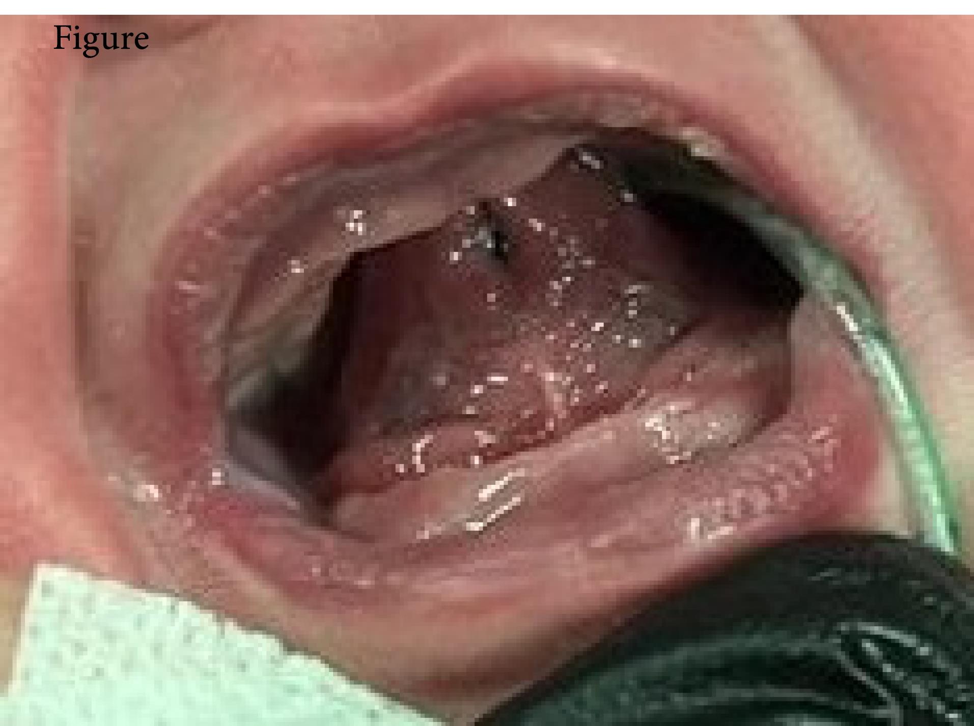
Figure 1.
Ankyloglossia superior: Tongue fused to the palate
.
Ankyloglossia superior: Tongue fused to the palate
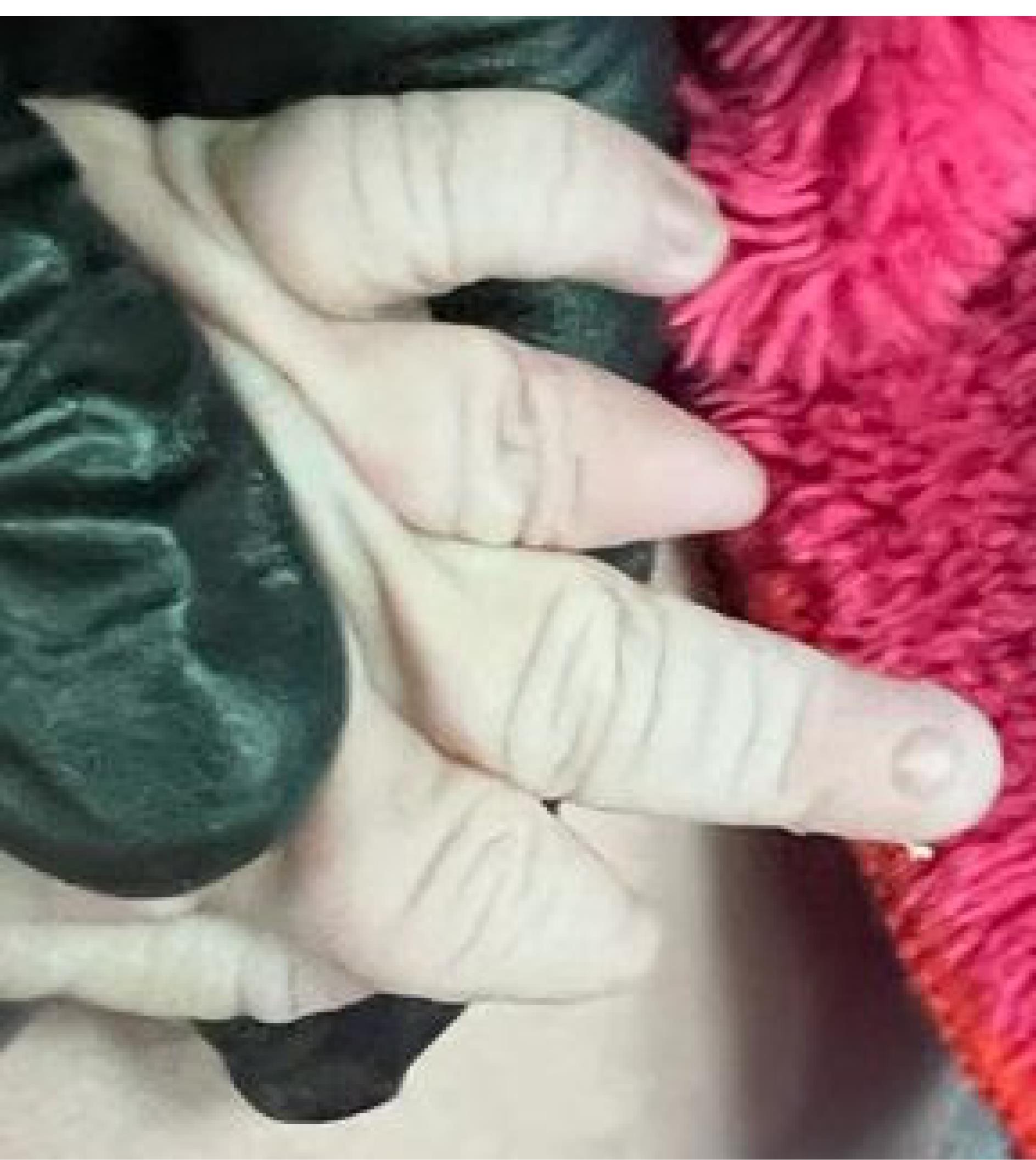
Figure 2.
The congenital lip deformity at the left hand
.
The congenital lip deformity at the left hand
Due to the presence of a large midline anterior mouth band, oral intubation was not possible. After that, we released the tissue linking the tongue to the hard palate under local anaesthetic (Figure 3). After removing fibrous bands, we discovered a bifid tongue and a wide cleft palate. Sutures were used to reconstruct the bifid tongue. After achieving hemostasis, 4-0 Vicryl sutures were used to enclose the wound edges. NGT was removed and mouth openness was improved. One day following the operation, the baby was spoon-fed. The procedure had no postoperative complications, and the patient was discharged from the hospital five days following the surgery with a satisfactory postoperative outcome. The patient had no trouble resuming oral intake at the postoperative checkup and showed no signs of misarticulation or dysarthria. The tongue mobility had changed by the time the patient underwent a second evaluation one month after the initial procedure. The cleft palate is corrected in 10 months after a monthly check-up with the patient.
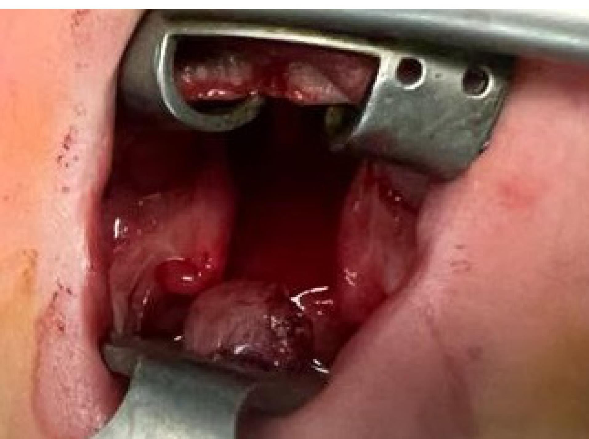
Figure 3.
After the tip of the tongue has been released
.
After the tip of the tongue has been released
Discussion
A disorder known as ankyloglossia is characterized by a short, thick lingual frenulum that attaches the tongue to the floor of the mouth, whereas superior ankyloglossia attaches the tongue’s tip to the palate. Superior ankyloglossia are related to aglossia-adactylia, hypodactylia, microglossia, Hanhart syndrome, syndactyly, Pierre Robin syndrome, ankyloglossia, Moebius syndrome, amniotic band syndrome, and Charlie M syndrome.2 Mandible, tongue, maxilla, and limbs are all affected by the aforementioned defects. Similar defects were superseded by the oromandibular limb hypogenesis syndrome, which were classified by Hall into five groups in 1971. The third group was assigned to superior ankyloglossia.3 Although superior ankyloglossia and aglossia adactylia share some characteristics, such as micrognathia hypoglossia or aglossia, cleft palates, or limb abnormalities, superior ankyloglossia can be distinguished from aglossia adactylia by the fibrotic band 135 years after Iller originally described the condition, the cause is still unclear. According to a number of theories about the pathogenesis of superior ankyloglossia, all anomalies occur during embryo development.2 Between the eighth and eleventh weeks of pregnancy, ankyloglossia superior syndrome is thought to have started to develop inside the uterus.4 At around the fourth week of pregnancy, the tongue starts to develop as two lateral lingual swellings and one medial swelling, tuberculum impar. The first pharyngeal arch is where the lingual swelling originates; in fact, the first pharyngeal arch is where the front two-thirds of the tongue is generated. The posterior of the tongue is made up of the second third, and a part of the fourth arch. Two shelf-like outgrowths from the maxillary protrusion that began growing in the seventh week make up the secondary palate. The secondary palate develops as the palatal shelves move from a vertical to a horizontal position above the tongue and fuse between weeks six and seven.5 Knowing how the mouth cavity develops could help pinpoint when superior ankyloglossia malformations first appeared. The pathophysiology of superior ankyloglossia has been the subject of several theories. A cleft palate and tongue adhesion to the palate can result from sedative medicines depressing the fetal swallowing reflex, which can also delay palatal shelf elevation, extend palatopharyngeal and palatoglossal epithelial contact, and delay palatal shelf elevation.6 According to the most widely accepted hypotheses, these bands are leftovers from the buccopharyngeal membrane. The thin membrane that divides the early pharynx from the mouth is called the buccopharyngeal membrane. The layer eventually disappears, but the tongue’s motility was constrained by the buccopharyngeal membrane, which remained2. The placement of the tongue between the palatal shelves prevents the fusion of the palatal shelves, which results in a cleft palate.7,8 According to another hypothesis, the adhesion between the tongue’s tip and the palate is caused by epithelial lesions of the tongue. The development of the tongue, palate, and limb was hampered by epithelial.9 According to another hypothesis, amniotic bands syndrome may be brought on by placental injury. Numerous congenital malformations, including limb or digital amputations, can result from the fiber band of the amniotic sac.10 Missense mutations in TX22 (the T.box transcription factor) can result in a cleft palate and ankyloglossia. The T.box gene is crucial for palatogenesis.11
Superior ankyloglossia may be caused by a variety of environmental and inherited variables.12 Urinary tract infections, drugs, vaginal hemorrhage, hypoxia, radiation, hypoglycemia, rubella, and avitaminosis are the examples of environmental and inherited variables that may be connected to superior ankyloglossia.2,13,14 In prior studies, 10 cases had cleft palates as a clinical characteristic, 13 had superior ankyloglossia with limb abnormalities, one had no further abnormalities, and two had congenital heart defects. In the majority of cases, general anesthesia was employed. Ours is the described example of superior ankyloglossia coupled with cleft palate, limb abnormalities, ASD, and tongue bifida (Table 1). One of the most difficult clinical scenarios is the problematic airway in the superior ankyloglossia because direct laryngoscopy is impossible due to the fibrous bands and direct laryngoscopy via endotracheal intubation is difficult and debatable. Numerous techniques have been employed to induce anesthesia in patients with superior ankyloglossia, including oral intubation using a paraglossal route, direct oral intubation, nasal intubation using a flexible pediatric bronchoscope, blind nasal intubation, and local anesthetic (Table 1), among them, we used local anesthesia in our case. A laryngeal mask is not ideal because it is easily misaligned.15
Table 1.
Summary of all reported cases of ankyloglossia superior syndrome
|
Gender
|
Age at operation
|
Limb anomalies
|
Cleft palate/defect
|
Other anomalies
|
Anesthea
|
Ref. |
| Male |
2days |
✓ |
✕ |
Tongue hypoplasia |
Not reported |
13
|
| Male |
Not reported |
✓ |
✕ |
Facial nerve paralysis, micrognathia, microglossia |
Local |
2
|
| Male |
Not reported |
✓ |
✓ |
GI anomaly, microglossia |
General |
14
|
| Female |
2 days |
✓ |
✕ |
✕ |
General |
16
|
| Male |
14 days |
✕ |
✓ |
✕ |
Local |
9
|
| Male |
1 year |
✓ |
✕ |
Microglossia, micrognathia |
General |
17
|
| Female |
5 months |
✓ |
✕ |
Micrognathia, hypoglossia |
General |
10
|
| Female |
45 days |
✕ |
✓ |
Strabismus, micrognathia |
General |
12
|
| Female |
2 days |
✕ |
✓ |
Bifid tongue |
Local |
18
|
| Male |
Not reported |
✕ |
✕ |
✕ |
Not reported |
19
|
| Female |
25 days |
✓ |
✕ |
Facial paralysis, GI, abnormality, abducens nerve palsy |
General |
20
|
| Male |
5 years |
✓ |
✓ |
✕ |
Local |
21
|
| Female |
Not reported |
✓ |
✓ |
✕ |
General |
15
|
| Male |
5 days |
✓ |
✕ |
Micrognathia |
General |
22
|
| Female |
0 days |
✓ |
✕ |
✕ |
Local |
23
|
| Male |
1 days |
✕ |
✓ |
Micrognathia, ASD, VSD |
General |
24
|
| Female |
5 months |
✓ |
✓ |
Micrognathia, moebius, syndrome, strabismus |
General |
25
|
| Female |
7 days |
✕ |
✓ |
Abnormal, genitalia, PDA |
General |
26
|
| Female |
42days |
✓ |
✓ |
Microglossia, natal teeth, micrognathia |
General |
27
|
| Female |
2 days |
✓ |
✓ |
ASD, bifid tongue |
Local |
Our case |
PDA: Patent Ductus Arteriosus; ASD: ankyloglossia superior syndrome
Surgery for superior ankyloglossia is scheduled based on the patient’s clinical status. In situations of elective surgery, the surgeon can hold off until the neonate has gained weight for a few weeks.16 Neonates with breathing or feeding issues require emergency surgery. However, difficulties with the temporomandibular joint result in prolonged surgery delays.
Conclusion
ASD is an uncommon congenital condition associated with craniofacial malformations, limb deformities, and gastrointestinal atresia. With initial multidisciplinary therapy, children with specific congenital abnormalities grow without substantial physical handicaps.
Competing Interests
The authors declare that they have no competing interests.
Consent for Publication
Written informed consent was obtained from the parents of patient to publish this case report and accompanying images.
Data Availability Statement
The datasets generated and analyzed during the current study are not publicly available due to legal reasons but are available from the corresponding author, H.S. on reasonable request.
Ethical Approval
Not Applicable.
Acknowledgements
We would like to appreciate the cooperation of Clinical Research Development Unit, Imam Reza General Hospital, Tabriz, Iran in conducting of this research.
References
- Illera MD. Congenital occlusion of the pharynx. Lancet 1887; 1:742-5. [ Google Scholar]
- Spivack J, Bennett JE. Glossopalatine ankylosis. Plast Reconstr Surg 1968; 42(2):129-36. doi: 10.1097/00006534-196808000-00005 [Crossref] [ Google Scholar]
- Hall BD. Aglossia-adactylia. Birth Defects Orig Artic Ser 1971; 7(7):233-6. [ Google Scholar]
- Gorlin RJ, Cohen MM Jr, Hennekam RC. Syndromes of the Head and Neck. Oxford University Press; 2001.
- Sadler TW. Langman’s Medical Embryology. Lippincott Williams & Wilkins; 2022.
- Humphrey T. The relation between human fetal mouth opening reflexes and closure of the palate. Am J Anat 1969; 125(3):317-44. doi: 10.1002/aja.1001250305 [Crossref] [ Google Scholar]
- Sybil D, Sagtani A. Cleft palate lateral synechia syndrome. Natl J Maxillofac Surg 2013; 4(1):87-9. doi: 10.4103/0975-5950.117876 [Crossref] [ Google Scholar]
- Gartlan MG, Davies J, Smith RJ. Congenital oral synechiae. Ann Otol Rhinol Laryngol 1993; 102(3):186-97. doi: 10.1177/000348949310200305 [Crossref] [ Google Scholar]
- Lekkas C, Bruaset I. Ankyloglossia superior. Oral Surg Oral Med Oral Pathol 1983; 55(6):556-7. doi: 10.1016/0030-4220(83)90368-7 [Crossref] [ Google Scholar]
- Gima H, Yamashiro M, Tomoyose Y. Ankyloglossum superius syndrome. J Oral Maxillofac Surg 1987; 45(2):158-60. doi: 10.1016/0278-2391(87)90406-x [Crossref] [ Google Scholar]
- Marçano AC, Doudney K, Braybrook C, Squires R, Patton MA, Lees MM. TBX22 mutations are a frequent cause of cleft palate. J Med Genet 2004; 41(1):68-74. doi: 10.1136/jmg.2003.010868 [Crossref] [ Google Scholar]
- Minami K, Sugahara T, Mori Y, Mishima K. Ankyloglossia superior: report of a case. J Oral Maxillofac Surg 1995; 53(5):588-9. doi: 10.1016/0278-2391(95)90073-x [Crossref] [ Google Scholar]
- Wilson RA, Kliman MR, Hardyment AF. Ankyloglossia superior (palato-glossal adhesion in the newborn infant). Pediatrics 1963; 31:1051-4. [ Google Scholar]
- Nevin NC, Kernohan DC, Ross AM. Ankyloglossum superius syndrome. Oral Surg Oral Med Oral Pathol 1980; 50(3):254-6. doi: 10.1016/0030-4220(80)90380-1 [Crossref] [ Google Scholar]
- Mukharjee S, Mitra D, Sen A, Chattopadhyay A, Kajal S, Dhankhar M. Intubation of a neonate with glossopalatine ankylosis using a paraglossal approach and a laryngoscope with a straight blade. South Afr J Anaesth Analg 2014; 20(5):36-7. doi: 10.10520/ejc163942 [Crossref] [ Google Scholar]
- Singh K, Sanasam Sanasam. Superior ankyloglossia. Indian J Otolaryngol 1981; 33(4):157. doi: 10.1007/bf02992294 [Crossref] [ Google Scholar]
- Chicarilli ZN, Polayes IM. Oromandibular limb hypogenesis syndromes. Plast Reconstr Surg 1985; 76(1):13-24. doi: 10.1097/00006534-198507000-00003 [Crossref] [ Google Scholar]
- Kalu PU, Moss AL. An unusual case of ankyloglossia superior. Br J Plast Surg 2004; 57(6):579-81. doi: 10.1016/j.bjps.2003.12.008 [Crossref] [ Google Scholar]
- Kothari PR, Gupta A. Ankyloglossia superior. Indian Pediatr 2005; 42(12):1249. [ Google Scholar]
- Bolling RP, Sabeeh V, Stewart JM Jr, Newsome RE, Chiu ES, Moses MH. Ankyloglossum superius syndrome: diagnosis and surgical management. J Craniofac Surg 2007; 18(5):1094-7. doi: 10.1097/scs.0b013e31814b2a0b [Crossref] [ Google Scholar]
- Wieker H, Sieg P. Ankyloglossia superior syndrome: case report and review of publications. Br J Oral Maxillofac Surg 2014; 52(5):464-6. doi: 10.1016/j.bjoms.2014.01.012 [Crossref] [ Google Scholar]
- Soe Khaing Thet Suu, Khin Maung1, Sun Sun Win. A case report of Superior Ankyloglossia. Myanmar Dental Journal 2015; 22(1):48-50. [ Google Scholar]
- Shay S, West AN. Ankyloglossia superior syndrome: case report and updated literature review. Int J Pediatr Otorhinolaryngol 2016; 86:1-3. doi: 10.1016/j.ijporl.2016.04.002 [Crossref] [ Google Scholar]
- Ohlstein JF, Padilla PL, Garza RK, Masel BD, Abouleish A, Pine HS. Ankyloglossum superius syndrome compromising a neonatal airway: considerations in congenital oral airway obstructions. Int J Pediatr Otorhinolaryngol 2019; 117:167-70. doi: 10.1016/j.ijporl.2018.11.036 [Crossref] [ Google Scholar]
- Freire RC, da Cunha Barbosa AP, Han MD, Borba AM. Ankyloglossia superior associated with moebius syndrome: a case report. J Oral Maxillofac Surg 2020; 78(1):87-90. doi: 10.1016/j.joms.2019.08.017 [Crossref] [ Google Scholar]
- Chowdhury TK, Chowdhury MZ, Sadia A, Farhad T, Alam MA. Superior ankyloglossia syndrome. J Pediatr Surg Case Rep 2020; 61:101611. doi: 10.1016/j.epsc.2020.101611 [Crossref] [ Google Scholar]
- Hara T, Tanaka S, Kogo M. Ankyloglossia superior syndrome with complex craniofacial anomalies: case report and literature review. Cleft Palate Craniofac J 2021; 58(7):906-11. doi: 10.1177/1055665620964028 [Crossref] [ Google Scholar]