J Res Clin Med. 13:35029.
doi: 10.34172/jrcm.025.35029
Systematic Review
Dendritic cell-derived extracellular vesicles as a promising immunotherapeutic strategy for cancer: A Systematic Review
Sahar Safaei Conceptualization, Data curation, Investigation, Writing – original draft, Writing – review & editing, 1, ## 
Sami Rassouli Conceptualization, 2, ##
Mahya Ahmadpour Youshanlui Project administration, Writing – review & editing, 1, #
Manouchehr Fadaee Methodology, 1, 3, #
Neda Shajari Data curation, Investigation, 1
Amin Khamaneh Methodology, 1
Behzad Baradaran Supervision, 1
Tohid Kazemi Supervision, 1, 3, *
Author information:
1Immunology Research Center, Tabriz University of Medical Sciences, Tabriz, Iran
2Student Research Committee, Tabriz University of Medical Sciences, Tabriz, Iran
3Department of Immunology, Faculty of Medicine, Tabriz University of Medical Sciences, Tabriz, Iran
#These authors contributed equally to this work.
##These two people share a first name.
Abstract
Introduction:
Despite advancements in cancer treatment, existing therapies like chemotherapy and radiotherapy often face challenges such as treatment resistance and adverse effects, necessitating the exploration of novel approaches. Dendritic cell-derived extracellular vesicles (DEVs) have emerged as a promising immunotherapeutic tool in cancer therapy. The purpose of this review is to summarize recent research on the immunotherapeutic potential of DEV, emphasizing their mechanism of action in the context of cancer treatment.
Methods:
Eligible studies were identified through comprehensive searches in PubMed, Scopus, and Google Scholar. Inclusion criteria encompassed original research on DEV’s immunotherapeutic efficacy, while reviews and non-original studies were excluded.
Results:
Nine studies were included, spanning from 2003 to 2022. The studies demonstrated that DEVs can activate CD8+cytotoxic T-cells, CD4+helper T-cells, and NK cells, thereby inducing potent antitumor immune responses. DEVs from interferon-treated dendritic cells exhibited enhanced antigen-presenting capabilities compared to other sources. DEVs also showed potential in regulating inflammation, with studies indicating suppression of the NF-κB signaling pathway in endothelial cells.
Conclusion:
DEVs represent a promising immunotherapeutic approach for cancer, capable of stimulating both innate and adaptive immune responses. However, challenges such as DEVs’ production variability and the need for standardized purification methods remain. Future research should focus on optimizing DEVs’ production and combining it with other immunotherapies to enhance therapeutic outcomes.
Keywords: Dendritic cells, Extracellular vesicles, Exosomes, Immunotherapy, Cancer
Copyright and License Information
© 2025 The Author(s).
This is an open access article distributed under the terms of the Creative Commons Attribution License (
http://creativecommons.org/licenses/by/4.0/), which permits unrestricted use, distribution, and reproduction in any medium, provided the original work is properly cited.
Funding Statement
None.
Introduction
The frequency of infections and the number of fatalities have significantly decreased as a result of vaccinations. Despite these advancements in medical research, effective solutions for cancer treatment remain elusive. In 2021, the World Health Organization recorded a significant 9.3 million deaths worldwide due to neoplasms, underscoring the urgent need for innovative cancer therapies.1,2
Complementary adjuvant chemotherapy and radiotherapy are essential components of comprehensive multidisciplinary cancer treatment strategies.3,4 However, resistance to radiotherapy and chemotherapy can lead to treatment inefficacy or cancer recurrence, complicating patient outcomes. While chemotherapy and radiotherapy are effective, their adverse effects limit their clinical use and often discourage patients from undergoing treatment. These limitations highlight the urgent necessity to explore novel cancer therapies that exhibit high specificity and efficacy in eliminating cancerous cells while preserving the integrity of healthy cells.5-9 The utilization of stem cells has garnered considerable attention in the field of cancer immunotherapy, primarily due to their capacity to differentiate into diverse immune cell populations.10,11 This innovative approach shows promise in overcoming challenges associated with existing immunotherapies, such as reduced tumor-killing potency and lack of in vivo specificity. Advancements in cell-based immunotherapy can be achieved by establishing cell lines that express tumor selectivity and cytotoxic functions.12,13 Extensive research has been dedicated to potential candidates, including dendritic cells (DCs), natural killer (NK) cells, macrophages, and T-lymphocytes, all of which hold immense promise in this field. These cells are essential in activating the immune system through chemotaxis, enhancing immune responses by triggering antibody-dependent and cytokine-dependent activation in other immune cells, and directly eliminating cancer cells through cytotoxic mechanisms.14,15 Their contributions are vital in orchestrating an effective immune response against cancer cells.
DC and CD4 + T-cells (T-helper cells) play crucial roles in regulating immune responses. They generate molecular signals such as cytokines and chemokines to guide and stimulate other immune cells.16,17 DCs are the most powerful antigen-presenting cells in the body, crucial for regulating immune responses and tolerance. They bridge innate and adaptive immunity by activating resting T cells. DCs express co-stimulatory molecules such as CD80 and CD86, which facilitate antigen uptake from tumors and the presentation of tumor-associated antigens (TAAs) via major histocompatibility complex (MHC) molecules.18,19
In addition to their antigen-presenting function, DCs secrete extracellular vehicles (EVs) known as DC-derived EVs (DEVs). DEVs are involved in immune responses and hold potential applications in cancer immunotherapies.20-22 DEVs carry TAAs and possess various molecules, including T-cell co-stimulatory molecules, antigen-presenting molecules, adhesion molecules, NK modulation molecules, and EV markers. The composition of DEVs depends on the physiological state of the DCs.23 DEVs have a significant role in activating T cells. They can directly present antigens to CD4 + and CD8 + T cells, leading to the suppression of tumor growth. DEVs can also be taken up by other DCs, providing exogenous peptide-loaded MHC (pMHC) for presentation to T cells.24 This process is relevant in organ transplantation, where recipient DCs incorporate donor DEVs to stimulate all specific T cells.25 DEVs can enhance T cell responses through a process called MHC cross-dressing, where they coat DCs with pMHC-loaded EVs. Additionally, DEVs can incorporate pMHC-loaded vesicles from other cell origins, such as epithelial cells, to enhance antigen presentation to T cells.26
Furthermore, DEVs can directly activate NK cells, inducing cytokine release and NK cell cytotoxicity through molecules like TNF-α. These immune responses, combined with apoptotic signaling, contribute to the removal of tumor cells 27. DEVs derived from heat-shocked-activated DCs carry BAT3, which acts as a ligand for NKp30 and mediates NK cell cytotoxicity.27 Conversely, tumor cells can secrete their EVs, known as tumor-derived EVs (TDEVs), which play a role in immune evasion. TDEVs can modulate the immune response by altering the tumor microenvironment and affecting various immune cells. They carry markers associated with tumor cells and can promote pro-inflammatory or anti-inflammatory responses depending on the target cells. TDEVs express programmed death-ligand 1 (PD-L1), which inhibits the functions of CD8 + T cells and contributes to adaptive immune resistance.28
Understanding the roles of these EVs provides valuable insights for the development of immunotherapies and cancer treatments. This review aims to synthesize current findings on the immunotherapeutic potential of DEVs, highlighting their mechanisms of action, therapeutic efficacy, and future directions in the context of cancer treatment.
Methods
Study design and search terms
This systematic review was conducted according to the guidelines set forth by the Preferred Reporting Items for Systematic Reviews and Meta-Analyses (PRISMA) statement. The primary aim was to assess the immunotherapeutic potential of DEVs in cancer treatment. The review involved comprehensive literature searches, study selection based on predefined criteria, risk of bias assessment, data extraction, and qualitative synthesis. Search terms included dendritic cell, extracellular vesicles, exosome, microvesicle, apoptotic body, tumor, cancer, and neoplasms.
Inclusion and exclusion criteria
Studies were included in the review if they involved DEVs used in cancer treatment, encompassing both experimental and clinical studies that reported on immunological outcomes, antitumor efficacy, or safety of DEVs. Eligible studies had to be original research articles available in English. Conversely, studies were excluded if they did not focus on cancer treatment, were reviews, editorials, or non-original research articles, lacked sufficient data on outcomes related to DEVs, or were not available in English.
Study selection
Initially, records were identified through database searches and manual searches of reference lists, with duplicates subsequently removed. In the screening stage, two independent reviewers assessed the titles and abstracts of the remaining records for relevance. During the eligibility assessment, full-text articles were evaluated to ensure they met the inclusion criteria by two independent authors. Any discrepancies between the reviewers were resolved through discussion or consultation with a third reviewer, providing a thorough and unbiased selection process.
Risk of bias assessment and data extraction
Risk of bias (RoB) assessments in the included clinical trials were conducted using the JBI critical appraisal tool for quasi-experimental studies. Data extraction was carried out independently by two reviewers using a standardized extraction form to ensure consistency and accuracy. Key data collected included study characteristics (author(s), year of publication, country, study design, and cell density), intervention details (source and type of DC, methods of DEV isolation and characterization, dosage, and administration routes), and outcomes (primary outcomes such as immune response and antitumor efficacy, and secondary outcomes like safety and side effects).
Data synthesis
Due to the heterogeneity in study designs, interventions, and outcome measures, data were synthesized qualitatively. A narrative synthesis approach was used to summarize the findings across studies, focusing on DEV’s mechanisms of action, immunotherapeutic potential, and clinical implications in cancer treatment.
Results
Results of the study selection
Nine studies were included in this review’s qualitative synthesis. These studies were selected based on their relevance to the immunotherapeutic use of DEVs in cancer treatment, providing comprehensive insights into their efficacy and potential as a novel cancer therapy. The PRISMA flow diagram illustrates the study selection process (Figure 1).
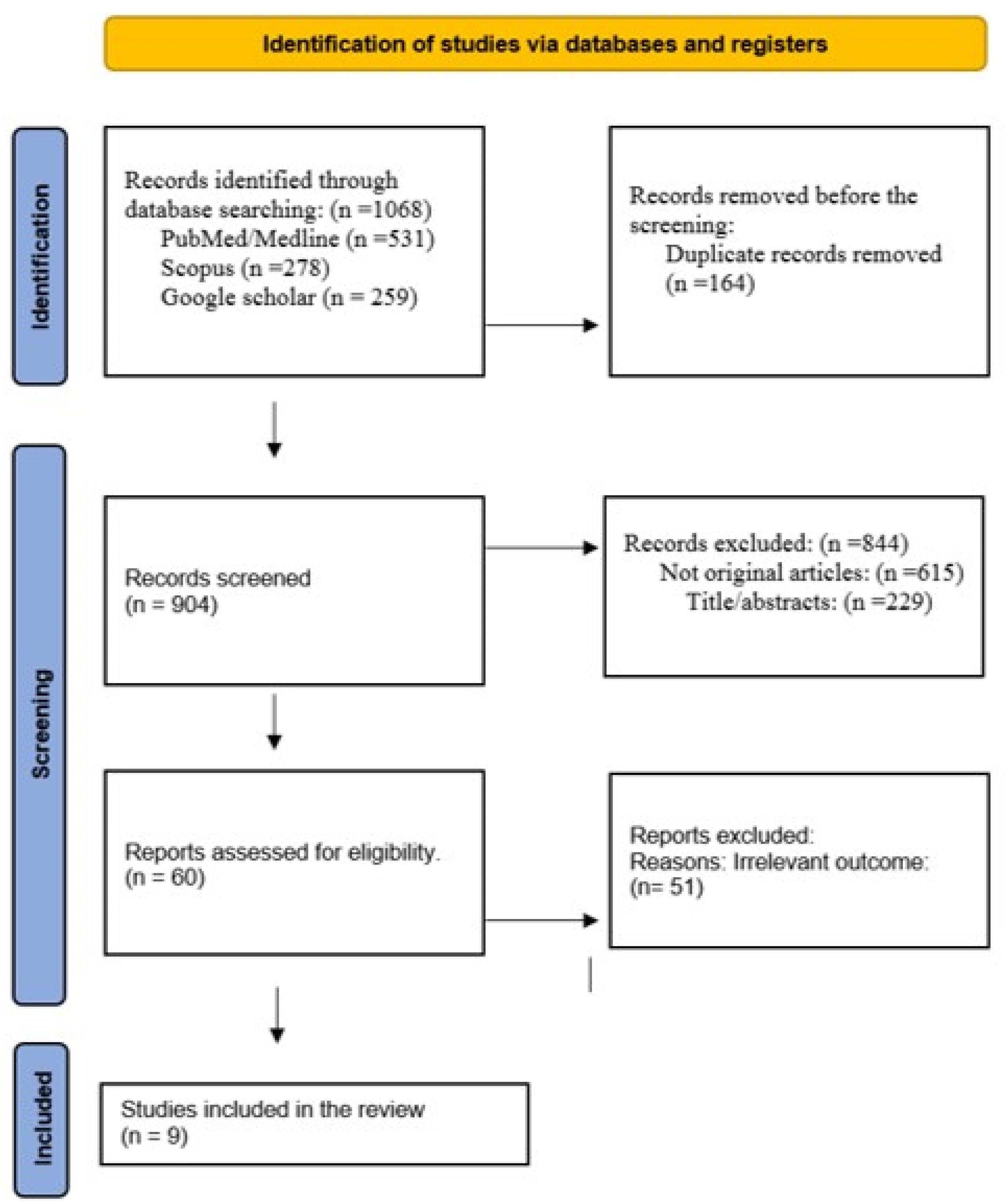
Figure 1.
PRISMA flow chart for systematic review
.
PRISMA flow chart for systematic review
After searching in (PubMed, Scopus, and Google Scholar) databases, a total of 1068 articles were obtained, and 164 duplicates were removed.
After reviewing the title & abstract screening, 60 studies remained. The final review includes nine articles of the final full-text results; the rest, which had unrelated data, were deleted.
Overview of included studies
The systematic review incorporated findings from nine studies published between 2003 and 2022, encompassing a global range of research from countries including France, China, Japan, the Netherlands, Vietnam, and Germany. Of these studies, two were clinical trials, and seven were experimental studies. The primary focus of these investigations was either the characterization of DEVs or their application in stimulating immune responses through pretreatment with immune system cells (Table 1).
Table 1.
Overview of studies on DEVs and immune response stimulation
|
Study
|
Year published
|
Original country
|
Study type
|
Target cells
|
| Clayton et al,29 |
2003 |
France |
Experimental |
B cell |
| Sakamato et al,30 |
2022 |
Japan |
Experimental |
T cell |
| Besse et al,31 |
2015 |
Germany |
Clinical Trial |
T and NK cell |
| Guan et al,32 |
2014 |
China |
Experimental |
T cell |
| Hsu et al,33 |
2003 |
France |
Experimental |
T cell |
| Li et al,34 |
2018 |
China |
Experimental |
T cell |
| Linderberg et al,35 |
2018 |
Netherlands |
Experimental |
T cell |
| Than et al,36 |
2020 |
Vietnam |
Experimental |
T cell |
| Viaud et al,37 |
2009 |
France |
Clinical Trial |
NK cell |
Purification and identification of exosomes
The predominant method for exosome purification was ultracentrifugation, with several studies also employing immunomagnetic capture and exosome precipitation kits. Exosome identification methods varied, including flow cytometry, transmission electron microscopy (TEM), and Western blotting. TEM was particularly noted for its effectiveness in directly detecting exosomes due to their small size (30-150 nm). Flow cytometry and immunofluorescence were useful for indirect identification when exosomes were coated with specific markers such as CD81 and CD63 (Table 2).
Table 2.
Method of purification and identification of DEVs
|
Study
|
Purification method
|
Identification of exosome
|
| Clayton et al,29 |
Ultracentrifugation Immunomagnetic
capture with anti-HLA-DP, -DQ, -DR |
Flow cytometry |
| Sakamato et al, 30 |
Exosome Precipitation Kit |
Flow cytometry |
| Besse et al,31 |
Ultracentrifugation |
Flow cytometry |
| Guan et al,32 |
Ultracentrifugation |
TEM, Western Blot |
| Hsu et al,33 |
Ultracentrifugation |
Flow cytometry |
| Li et al,34 |
Exosome Precipitation Kit |
TEM, Western Blot |
| Lindenbergh et al,35 |
Ultracentrifugation |
Western Blot |
| Than et al,36 |
Ultracentrifugation |
TEM, Western Blot |
| Viaud et al,37 |
Ultracentrifugation |
Flow cytometry |
TEM, transmission electron microscopy.
Immunotherapeutic potential and functional analysis
DEVs have shown significant promise as an immunotherapeutic agent through various mechanisms (Table 3).
Table 3.
In vitro evidence demonstrating the immunotherapeutic potential of DC-EVs
|
Study
|
Type of NK-EV
|
Cell density NK-EV
|
Methods of assessment
|
Key findings
|
| Clyton et al,29 |
PBMC of healthy people |
1 × 10⁶ cells |
Complement membrane regulator proteins |
The APC-derived exosomes express CD55 and CD59 proteins but not CD46 GPI-anchored complement regulators are expressed by exosomes to enable their viability in the extracellular space. |
| Sakamato et al,30 |
MUTZ-3 cells |
1 × 10⁵ cells |
Expression levels of surface molecules, such as CD80, CD86, CD83, HLA-ABC, HLA-DR and CD40 for antigen presentation ability |
MUTZ-3-derived DCs can produce distinct dexosomes with different phenotypes, and in particular, dexosomes derived from IFN-induced DCs exhibit higher expression levels of HLA-ABC compared to those derived from IL-4-induced DCs
Dexosomes derived from IFN-induced DCs (M-mIFN-Dex) have a greater ability to present antigens to CD8 + T cells compared to dexosomes derived from IL-4-induced DCs (M-mIL-4-Dex) |
| Besse et al,31 |
MD-DC from HLA-A2 + healthy donor |
0.13 × 10¹⁴ cells
four intradermal exosome vaccinations at one-week intervals |
T cell immune response and NK cell function |
IFN-γ- DEVs did not induce cancer-specific T-cell immune responses. IFN-γ- DEVs increases NKp30-related NK cell functions. Lack of control group was a possible source of bias in this study. |
| Guan et al,32 |
Umbilical cord blood-derived DC |
1 μg, 2 μg, 5 μg, 10 μg, 20 μg |
Stimulation of T lymphocytes |
DEVs can increase T cell proliferation, cytotoxic effects, and improve in vivo anticancer immunity. |
| Hsu et al,33 |
Monocyte-derived from a healthy donor |
100 μL |
Stimulation of T lymphocytes |
Exosomes can activate CD4 + T helper cells and CD8 + cytotoxic T cells, both of which are essential for a potent antitumor response. |
| Li et al,34 |
BMDCs derived exosomes |
25 μL, 50 μL, and 100 μL |
Effect on TNF-α–induced endothelial inflammation. |
Exosomes obtained from BMDCs treated with LIPUS inhibit the inflammatory response of endothelial cells induced by TNFα by suppressing the NF-κB signaling pathway. |
| Lindenbergh et al, 35 |
LPS-activated autologous moDC derived-exosomes (from healthy donors) |
NR |
Stimulating
peptide-specific CD8 + T-cells |
Monocyte-derived DCs increased the release of specific proteins and microRNAs, with immune-stimulatory capacities, in their EVs in response to interaction with activated bystander T-cells, and these EVs are functionally active in stimulating peptide-specific CD8 + T-cells. |
| Than et al,36 |
Cryopreserved umbilical cord blood mononuclear cell-derived dcs (cryo cbmdcs) |
6 × 10⁵ cells/100 µL) |
CD3 + T cells from healthy donors’ PBMCs |
Immune cells induced by DEVs showed greater cytotoxicity against A549 tumor cells |
| Viaud et al,37 |
DC derived-exosomes |
10 μg |
NK cell activation
-Phase I trial: Autologous NK cells and CD8 + T cells from melanoma patients. |
Human DEVs expresses functional IL-15Rα molecules, which could trans-present exogenous IL-15 to NK cells, enhancing the NK cell proliferation and IFNγ production in vitro. Human DEVs can activate NK cells through an NKG2D-dependent mechanism, similar to mouse DEVs. Lack of control group was a possible source of bias in this study. |
PBMC, peripheral blood mononuclear cells; DEVs, dendritic cell-derived extracellular vesicles; BMDCs, bone marrow-derived cells; LIPUS, low-intensity pulsed ultrasonography; TNF, tumor necrosis factor; IL, interleukin
Activation of T cells: Multiple studies have demonstrated DEVs’ ability to stimulate both CD8 + cytotoxic T cells and CD4 + helper T cells, which is essential for potent antitumor responses. For example, Guan et al32 reported that umbilical cord blood-derived DEVs increased T cell proliferation and cytotoxic effects, enhancing anticancer immunity in vivo. Similarly, Hsu et al33 found that DEVs activated CD4 + and CD8 + T cells, contributing to a robust antitumor response.
NK cell stimulation: DEVs also played a crucial role in enhancing NK cell function. Besse et al31 observed that DEVs increased NKp30-related NK cell functions, although they did not induce cancer-specific T cell immune responses. Viaud et al37 demonstrated that human DEVs could activate NK cells through a mechanism dependent on NKG2D ligands and interleukin (IL)-15Rα, leading to increased NK cell proliferation and IFN-γ production.
Cytotoxicity against tumor cells: Than et al36 showed that immune cells primed with DEVs exhibited increased cytotoxicity against A549 tumor cells, highlighting the potential of DEVs to bridge natural and antigen-specific T cell responses and contribute to tumor regression.
Regulation of inflammatory responses: Li et al34 found that exosomes from bone marrow-derived DCs treated with low-intensity pulsed ultrasonography could inhibit inflammatory responses in endothelial cells by suppressing the NF-κB signaling pathway, indicating a potential for DEVs in modulating inflammation-related aspects of cancer progression.
Exosome biomarkers and functional attributes
Studies also focused on the specific biomarkers and functions of DEVs:
Complement membrane regulator proteins: Clayton et al29 identified that exosomes expressed CD55 and CD59 but not CD46, which are essential for maintaining exosome viability in the extracellular space.
Antigen presentation: Sakamoto et al30 showed that DEVs from IFN-induced DCs had a greater ability to present antigens to CD8 + T cells compared to those from IL-4-induced DCs.
Immune cell priming: Lindenbergh et al35 noted that exosomes from LPS-activated autologous monocyte-derived DCs increased the release of specific proteins and microRNAs with immune-stimulatory capacities, enhancing peptide-specific CD8 + T cell responses.
Discussion
This systematic review comprehensively evaluated the potential of DEVs as immunotherapeutic agents for cancer treatment. The nine studies included in this review, spanning from 2003 to 2022, elucidated various aspects of DEVs’ biological functions, their mechanisms of action, and their therapeutic implications. The key findings reveal that DEVs are capable of activating both T cells and NK cells, thereby inducing potent antitumor immune responses. Additionally, the studies underscored the effectiveness of ultracentrifugation and other advanced methods for exosome purification and characterization.
The ability of DEVs to activate CD4 + and CD8 + T cells is a critical finding, as these cells are essential for a robust and sustained antitumor response. Guan et al32 and Hsu et al33 demonstrated the enhanced proliferation and cytotoxicity of T cells when stimulated by DEVs, corroborating the potential of these vesicles in eliciting adaptive immune responses. Furthermore, the activation of NK cells, as shown by Besse et al31 and Viaud et al,37 highlights another crucial aspect of DEVs’ functionality, emphasizing their role in innate immunity. DEVs’ dual activation of both arms of the immune system suggests a comprehensive approach to immunotherapy, which is likely to result in more effective tumor eradication.
Clinical application of cell-based therapies appears challenging but promising.38 Clinical trials included in the review demonstrated the feasibility and potential efficacy of DEVs-based treatments in human patients. For instance, Viaud et al37 and Besse et al31 conducted trials that showed enhanced NK cell functions and T cell responses, albeit with varying degrees of success in eliciting cancer-specific responses. These trials underscore the necessity for further optimization and standardization of DEVs production and administration protocols to maximize therapeutic outcomes.
Despite the promising results, several challenges remain. One significant issue is the variability in DEVs’ production and purification methods. Ultracentrifugation, although widely used, may not consistently yield high-purity exosome preparations, which can affect the reproducibility and efficacy of DEVs-based therapies. Future research should focus on refining these techniques, possibly incorporating more advanced and standardized methods such as immunomagnetic capture and exosome precipitation kits, as demonstrated by Sakamoto et al30 and Li et al.34
Another challenge is DEVs heterogeneity of DEVs. The source of DCs and the conditions under which they are cultured can significantly influence the composition and functional properties of DEVs. This review highlights the need for a deeper understanding of these variables to ensure DEVs’ preparations are consistently potent and effective. For example, Sakamoto et al30 showed that DEVs derived from IFN-induced DCs had superior antigen-presenting capabilities compared to those from IL-4-induced cells, indicating the importance of the DC maturation state in DEVs’ efficacy.
Future studies should also explore the integration of DEVs with other immunotherapeutic strategies, such as checkpoint inhibitors and adoptive cell transfer, to enhance their antitumor effects. The combination of DEVs with these therapies could overcome some of the limitations observed in monotherapy trials, leading to more robust and sustained responses.
Conclusion
The findings from this systematic review underscore the substantial potential of DEVs as a novel immunotherapeutic strategy for cancer. DEVs’ ability to activate both T cells and NK cells positions it as a powerful agent in the fight against cancer. However, further research is necessary to address the challenges of production standardization, heterogeneity, and clinical application. With continued advancements in the field, DEVs-based therapies hold promise for becoming a cornerstone of cancer immunotherapy, offering new hope for patients with challenging malignancies.
Competing Interests
The authors declare that they have no known competing financial interests or personal relationships that could have appeared to influence the work reported in this paper.
Ethical Approval
Ethical approval and informed consent were not required for this study, as these studies were based on previously published articles.
References
- Manshouri R, Coyaud E, Kundu ST, Peng DH, Stratton SA, Alton K. ZEB1/NuRD complex suppresses TBC1D2b to stimulate E-cadherin internalization and promote metastasis in lung cancer. Nat Commun 2019; 10(1):5125. doi: 10.1038/s41467-019-12832-z [Crossref] [ Google Scholar]
- Safaei S, Fadaee M, Rahbar Farzam O, Yari A, Poursaei E, Aslan C. Exploring the dynamic interplay between exosomes and the immune tumor microenvironment: implications for breast cancer progression and therapeutic strategies. Breast Cancer Res 2024; 26(1):57. doi: 10.1186/s13058-024-01810-z [Crossref] [ Google Scholar]
- van Velsen EF, Stegenga MT, van Kemenade FJ, Kam BL, van Ginhoven TM, Visser WE. Evaluation of the 2015 ATA guidelines in patients with distant metastatic differentiated thyroid cancer. J Clin Endocrinol Metab 2020; 105(3):e457-65. doi: 10.1210/clinem/dgz137 [Crossref] [ Google Scholar]
- Jafari F, Javdansirat S, Sanaie S, Naseri A, Shamekh A, Rostamzadeh D. Osteosarcoma: a comprehensive review of management and treatment strategies. Ann Diagn Pathol 2020; 49:151654. doi: 10.1016/j.anndiagpath.2020.151654 [Crossref] [ Google Scholar]
- Zhang QY, Wang FX, Jia KK, Kong LD. Natural product interventions for chemotherapy and radiotherapy-induced side effects. Front Pharmacol 2018; 9:1253. doi: 10.3389/fphar.2018.01253 [Crossref] [ Google Scholar]
- Safaei S, Amini M, Najjary S, Mokhtarzadeh A, Bolandi N, Saeedi H. miR-200c increases the sensitivity of breast cancer cells to doxorubicin through downregulating MDR1 gene. Exp Mol Pathol 2022; 125:104753. doi: 10.1016/j.yexmp.2022.104753 [Crossref] [ Google Scholar]
- Daei Sorkhabi A, ZareDini M, Fazlollahi A, Sarkesh A, Naseri A, Mousavi SE. The safety and efficacy of tislelizumab, alone or in combination with chemotherapy, for the treatment of non-small cell lung cancer: a systematic review of clinical trials. BMC Pulm Med 2023; 23(1):495. doi: 10.1186/s12890-023-02755-3 [Crossref] [ Google Scholar]
- Tirabadi S, Nahai MR, Alizadeh N, Ahmadpour Youshanlui M, Baradaran B. Different concentrations of produced recombinant TNF-α protein caused different cellular viability in breast cancer cell lines. Int J Drug Res Clin 2024; 2(1):e18. doi: 10.34172/ijdrc.2024.e18 [Crossref] [ Google Scholar]
- Abolhasani S, Javadi S, Hassannia H, Ghalamfarsa G, Hojjat-Farsangi M, Jadidi-Niaragh F. IGF1R and HIF-1α gene silencing inhibits cancer cell growth. Int J Drug Res Clin 2023; 1(1):e17. doi: 10.34172/ijdrc.2023.e17 [Crossref] [ Google Scholar]
- Ren J, Zhang X, Zhang Z, Pan J, Hao Z, Li J. Apoptosis inhibition enhances induced pluripotent stem cell generation during T cell reprogramming. Biochem Biophys Res Commun 2023; 656:30-7. doi: 10.1016/j.bbrc.2023.03.024 [Crossref] [ Google Scholar]
- Feizi H, Hosseini MS, Seyedi-Sahebari S, Karimi H, Mosaddeghi-Heris R, Sadigh-Eteghad S. A systematic review of clinical efficacy and safety of cell-based therapies in Alzheimer’s disease. Dement Neuropsychol 2024; 18:e20240147. doi: 10.1590/1980-5764-dn-2024-0147 [Crossref] [ Google Scholar]
- Wang Y, Wang C, Liu T, Qi H, Chen S, Cai X. Discovery of tetrahydropyrazolopyrazine derivatives as potent and selective MYT1 inhibitors for the treatment of cancer. J Med Chem 2024; 67(1):420-32. doi: 10.1021/acs.jmedchem.3c01476 [Crossref] [ Google Scholar]
- Farajzadeh M, Hosseini MS, Eshaghi F, Mohebbi S, Hojjat-Farsangi M, Jadidi-Niaragh F. Combined blockade of interleukin-6 and β-catenin inhibits breast and colon tumor growth in vitro. Int J Drug Res Clin 2024; 2(1):e19. doi: 10.34172/ijdrc.2024.e19 [Crossref] [ Google Scholar]
- Parikh BA, Piersma SJ, Pak-Wittel MA, Yang L, Schreiber RD, Yokoyama WM. Dual requirement of cytokine and activation receptor triggering for cytotoxic control of murine cytomegalovirus by NK cells. PLoS Pathog 2015; 11(12):e1005323. doi: 10.1371/journal.ppat.1005323 [Crossref] [ Google Scholar]
- Masoumi J, Ghorbaninezhad F, Saeedi H, Safaei S, Khaze Shahgoli V, Ghaffari Jolfayi A. siRNA-mediated B7H7 knockdown in gastric cancer lysate-loaded dendritic cells amplifies expansion and cytokine secretion of autologous T cells. Biomedicines 2023; 11(12):3212. doi: 10.3390/biomedicines11123212 [Crossref] [ Google Scholar]
- Hilligan KL, Ronchese F. Antigen presentation by dendritic cells and their instruction of CD4 + T helper cell responses. Cell Mol Immunol 2020; 17(6):587-99. doi: 10.1038/s41423-020-0465-0 [Crossref] [ Google Scholar]
- Ghorbaninezhad F, Asadzadeh Z, Masoumi J, Mokhtarzadeh A, Kazemi T, Aghebati-Maleki L. Dendritic cell-based cancer immunotherapy in the era of immune checkpoint inhibitors: From bench to bedside. Life Sci 2022; 297:120466. doi: 10.1016/j.lfs.2022.120466 [Crossref] [ Google Scholar]
- Qian C, Cao X. Dendritic cells in the regulation of immunity and inflammation. Semin Immunol 2018; 35:3-11. doi: 10.1016/j.smim.2017.12.002 [Crossref] [ Google Scholar]
- Moussion C, Delamarre L. Antigen cross-presentation by dendritic cells: a critical axis in cancer immunotherapy. Semin Immunol 2024; 71:101848. doi: 10.1016/j.smim.2023.101848 [Crossref] [ Google Scholar]
- Kowal J, Tkach M. Dendritic cell extracellular vesicles. Int Rev Cell Mol Biol 2019; 349:213-49. doi: 10.1016/bs.ircmb.2019.08.005 [Crossref] [ Google Scholar]
- Schierer S, Ostalecki C, Zinser E, Lamprecht R, Plosnita B, Stich L. Extracellular vesicles from mature dendritic cells (DC) differentiate monocytes into immature DC. Life Sci Alliance 2018; 1(6):e201800093. doi: 10.26508/lsa.201800093 [Crossref] [ Google Scholar]
- Li J, Li J, Peng Y, Du Y, Yang Z, Qi X. Dendritic cell derived exosomes loaded neoantigens for personalized cancer immunotherapies. J Control Release 2023; 353:423-33. doi: 10.1016/j.jconrel.2022.11.053 [Crossref] [ Google Scholar]
- Fernández-Delgado I, Calzada-Fraile D, Sánchez-Madrid F. Immune regulation by dendritic cell extracellular vesicles in cancer immunotherapy and vaccines. Cancers (Basel) 2020; 12(12):3558. doi: 10.3390/cancers12123558 [Crossref] [ Google Scholar]
- Speiser DE, Chijioke O, Schaeuble K, Münz C. CD4 + T cells in cancer. Nat Cancer 2023; 4(3):317-29. doi: 10.1038/s43018-023-00521-2 [Crossref] [ Google Scholar]
- Fortunato M, Morali K, Passeri L, Gregori S. Regulatory cell therapy in organ transplantation: achievements and open questions. Front Immunol 2021; 12:641596. doi: 10.3389/fimmu.2021.641596 [Crossref] [ Google Scholar]
- Chatterjee F, Spranger S. MHC-dressing on dendritic cells: Boosting anti-tumor immunity via unconventional tumor antigen presentation. Semin Immunol 2023; 66:101710. doi: 10.1016/j.smim.2023.101710 [Crossref] [ Google Scholar]
- Wei L, Xiang Z, Zou Y. The role of NKG2D and its ligands in autoimmune diseases: new targets for immunotherapy. Int J Mol Sci 2023; 24(24):17545. doi: 10.3390/ijms242417545 [Crossref] [ Google Scholar]
- Wang S, Sun J, Dastgheyb RM, Li Z. Tumor-derived extracellular vesicles modulate innate immune responses to affect tumor progression. Front Immunol 2022; 13:1045624. doi: 10.3389/fimmu.2022.1045624 [Crossref] [ Google Scholar]
- Clayton A, Harris CL, Court J, Mason MD, Morgan BP. Antigen-presenting cell exosomes are protected from complement-mediated lysis by expression of CD55 and CD59. Eur J Immunol 2003; 33(2):522-31. doi: 10.1002/immu.200310028 [Crossref] [ Google Scholar]
- Sakamoto T, Koya T, Togi M, Yoshida K, Kato T Jr, Ishigaki Y. Different in vitro-generated MUTZ-3-derived dendritic cell types secrete dexosomes with distinct phenotypes and antigen presentation potencies. Int J Mol Sci 2022; 23(15):8362. doi: 10.3390/ijms23158362 [Crossref] [ Google Scholar]
- Besse B, Charrier M, Lapierre V, Dansin E, Lantz O, Planchard D. Dendritic cell-derived exosomes as maintenance immunotherapy after first line chemotherapy in NSCLC. Oncoimmunology 2016; 5(4):e1071008. doi: 10.1080/2162402x.2015.1071008 [Crossref] [ Google Scholar]
- Guan S, Li Q, Liu P, Xuan X, Du Y. Umbilical cord blood-derived dendritic cells loaded with BGC823 tumor antigens and DC-derived exosomes stimulate efficient cytotoxic T-lymphocyte responses and antitumor immunity in vitro and in vivo. Cent Eur J Immunol 2014; 39(2):142-51. doi: 10.5114/ceji.2014.43713 [Crossref] [ Google Scholar]
- Hsu DH, Paz P, Villaflor G, Rivas A, Mehta-Damani A, Angevin E. Exosomes as a tumor vaccine: enhancing potency through direct loading of antigenic peptides. J Immunother 2003; 26(5):440-50. doi: 10.1097/00002371-200309000-00007 [Crossref] [ Google Scholar]
- Li X, Li X, Lin J, Sun X, Ding Q. Exosomes derived from low-intensity pulsed ultrasound-treated dendritic cells suppress tumor necrosis factor-induced endothelial inflammation. J Ultrasound Med 2019; 38(8):2081-91. doi: 10.1002/jum.14898 [Crossref] [ Google Scholar]
- Lindenbergh MF, Koerhuis DG, Borg EG, van ‘t Veld EM, Driedonks TA, Wubbolts R. Bystander T-cells support clonal T-cell activation by controlling the release of dendritic cell-derived immune-stimulatory extracellular vesicles. Front Immunol 2019; 10:448. doi: 10.3389/fimmu.2019.00448 [Crossref] [ Google Scholar]
- Than UT, Le HT, Hoang DH, Nguyen XH, Pham CT, Bui KT. Induction of antitumor immunity by exosomes isolated from cryopreserved cord blood monocyte-derived dendritic cells. Int J Mol Sci 2020; 21(5):1834. doi: 10.3390/ijms21051834 [Crossref] [ Google Scholar]
- Viaud S, Terme M, Flament C, Taieb J, André F, Novault S. Dendritic cell-derived exosomes promote natural killer cell activation and proliferation: a role for NKG2D ligands and IL-15Ralpha. PLoS One 2009; 4(3):e4942. doi: 10.1371/journal.pone.0004942 [Crossref] [ Google Scholar]
- Salehi-Pourmehr H, Nouri O, Naseri A, Roshangar L, Rahbarghazi R, Sadigh-Eteghad S. Clinical application of stem cell therapy in neurogenic bladder: a systematic review and meta-analysis. Int Urogynecol J 2022; 33(8):2081-97. doi: 10.1007/s00192-021-04986-6 [Crossref] [ Google Scholar]